For Which of the Following Is Bitewing Film Used
This in turn signifies less radiation exposure to the patient and less time spent with uncomfortable sensor equipment in his or her mouth. This brings the total cost of a bitewing series of four periapical films to 129 cents assuming that a new bitewing tab is used for each periapical films as compared to 148 cents for two regular bite- wing films.

Bitewing Technique L Oral Radiology Mcqs Wikidentia
Left molar bitewing film was bent when it was placed into the image receptor holder.
. Bone about 2 to 3 mm beyond the apex. If the size 2 film sensor is being used the anterior films are in the _____ position and the posterior filmssensors are in the _____ position. When the film is parallel with the long axis of the tooth the image looks the same as the tooth itself.
Fixed solution was low. A film is inserted between the cardboard and the whole device is placed in the patient mouth. Radiographic x-ray imaging is an integral tool in the assessment and diagnosis of dental caries tooth decay and periodontal gum disease.
The most radiolucent area on an anterior radiograph is the. An intraoral full-mouth survey FMX on an adult consist of --------- images. Mandibular incisors periapical radiograph appears very light with a hint of a diamond-like pattern superimposed over the image of the teeth.
A periapical view shows the tooth from the occlusal surface or incisal edge to the. A full mouth series of periapical and bitewing radiographs exhibits the following errors. The intraoral film used to show desired teeth their apices and surrounding areas are called ____ films.
It is having a holder which is made-up of cardboard. Class II dental malocclusion in the mixed dentition will most likely Proper collimation of the useful beam for the film size and target-film distance. There are four sizes of Bitewing radiographic films based on the position and the age of the patient it is used in.
A bitewing radiograph of an early mixed dentition should include the following proximal surfaces. To present a complete image in the. Horizontal Bitewing Film Orientation.
Good contrast and density Noimage. In this study in order to separate each tooth in the bitewing image first horizontal projection was applied to separate the upper and lower rows of teeth in the bitewing film into two photos while vertical projection was applied to separate the individual teeth from the upper and lower rows of teeth into a single individual tooth photo. These are great for horizontal bitewings and allow you to line up the collimator well.
Our study compared the diagnostic quality of bitewing radiographs of. Tumour risk of dental x-rays. Thus the four-film series is 18 cents less expensive per patient than the regular two-film bitewing series.
Terms in this set 18 The bitewing view is used for detecting. Evidence-based research clearly shows that used judiciously bitewing x-ray imaging is the method of choice for determining the presence and extent of decay in the areas where back. It is a film used in dental to show the crowns of upper and lower teeth simultaneously.
Interproximal decay and periodontal disease. A form of entertainment information etc composed of such a sequence of images and shown in a cinema etc. Film The paralleling technique is the preferred method and will be illustrated throughout this chapter.
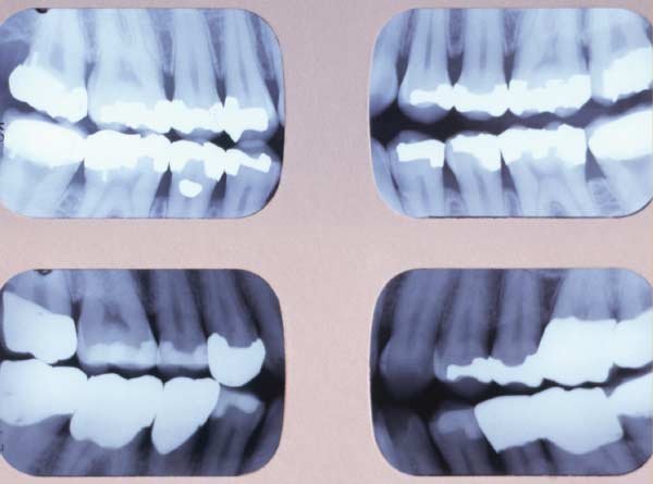
Like photographic film the faster the film the less exposure. Routine bitewing radiographs are commonly used to examine for interdental caries and recurrent caries under. Diagnostic images show the following characteristics.
Dear Bill Lets start with the bottom line. If your patient cant manage with the film holders or you want to try vertical bitewings I have found the following to be really useful. Film-holding devices have been recommended as a means of improving the diagnostic yield.
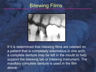
The bitewing view is taken to visualize the crowns of the posterior teeth and the height of the alveolar bone in relation to the cementoenamel junctions which are the demarcation lines on the teeth which separate tooth crown from tooth root. Determine if the following basic principles of the bitewing technique are true or false. When taking mandibular and bitewing films the dental assistant must have the patient lift her ____ so that the lower border of the film packet is directly lingual.
As modifier. There is no distortion. As the name suggests this films is used to record the Maxilla or Mandible from the occlusal surface showing all the teeth Oclussaly.
Bitewing digital x-rays require fewer retakes as a result of under- or overexposure or positioning errors both problems often associated with conventional film x-ray technology. The following items should be removed from the patient. This type of Radiographic technique is used in recording the position and number of Supernumerary or Impacted teeth in.
A sequence of images of moving objects photographed by a camera and providing the optical illusion of continuous movement when projected onto a screen. Cells most susceptible to radiation damage are. The film typically used for the intraoral bitewing exam falls into three film speed classes - D slowest E and F-speed fastest.
Which of the following bitewing film size is the longest. Which of the following could cause a straight black lineborder to appear on the radiograph. One of the most common procedures is the bitewing X-ray which uses an X-ray film clenched between the teeth in a tab of plastic or cardboard to check for decay between the teeth.

Examples Of Bitewing Films With The Correct Number Of Teeth Download Scientific Diagram
Radiology Bitewing Technique
Bitewing X Rays


Comments
Post a Comment